File:PET-schema.png
Jump to navigation
Jump to search

Size of this preview: 800 × 586 pixels. Other resolutions: 320 × 235 pixels | 640 × 469 pixels | 1,024 × 750 pixels | 1,280 × 938 pixels.
Original file (1,280 × 938 pixels, file size: 698 KB, MIME type: image/png)
Summary
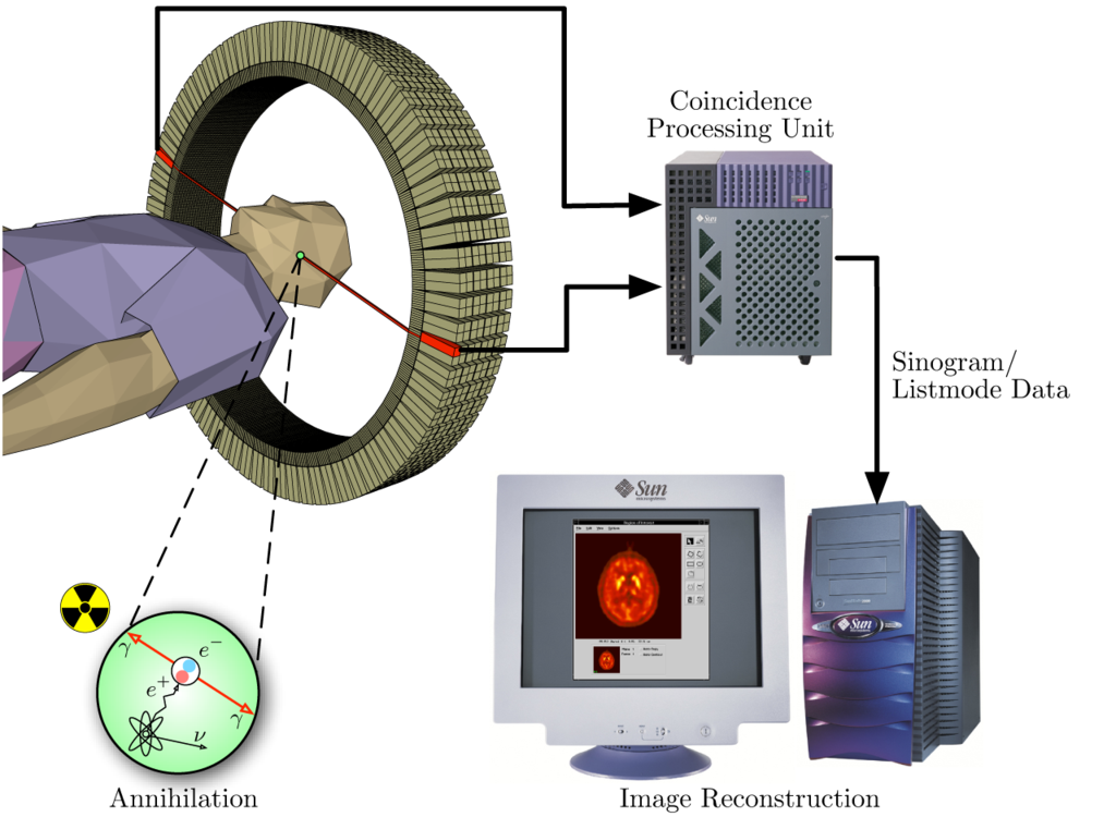
| Description | The image illustrates the processing principles of a positron emission tomograph (PET) commonly used in cancer diagnostics. It shows how during the annihilation process two photons are emitted in diametrically opposite directions. These photons are registered by the scanner as soon as they arrive at the detector ring. After the registration, the data is forwarded to a processing unit which decides if two registered events are selected as a so-called coincidence event. All coincidences are forwarded to the image processing unit where the final image data is produced via mathematical image reconstruction procedures. | ||
| Date | |||
| Source |
own work - part of master thesis |
||
| Author | Jens Maus (http://jens-maus.de/) | ||
| Permission (Reusing this file) |
|
File history
Click on a date/time to view the file as it appeared at that time.
| Date/Time | Thumbnail | Dimensions | User | Comment | |
|---|---|---|---|---|---|
| current | 17:25, 17 November 2005 | | 1,280 × 938 (698 KB) | wikimediacommons>Damato | Uploaded a version with a higher resolution. Content not changed. |
File usage
There are no pages that use this file.